|
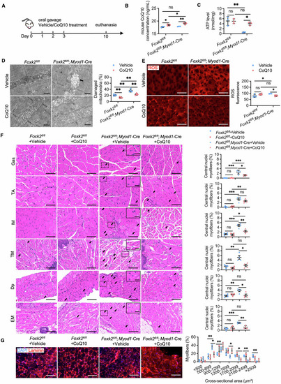
Coenzyme Q10 improves mitochondrial dysfunction and skeletal muscle development disorders of mice with Foxk2 deficiency in muscle stem cells. (A) Illustration of the study design of CoQ10 treatment in mice. (B) Concentration of CoQ10 in TA tissues of mice at 2 weeks (n = 3 mice). P value: **P = 0.0013; ns-P = 0.4524; Foxk2fl/fl+Vehicle vs Foxk2fl/fl + CoQ10, *P = 0.0118; Foxk2fl/fl + CoQ10 vs Foxk2fl/fl;Myod1-Cre+CoQ10, *P = 0.0435. (C) Quantification of ATP content in TA tissues from Foxk2fl/fl and Foxk2fl/fl; Myod1-Cre littermates at 2 weeks, with or without CoQ10 treatment (n = 3 mice). Values were normalized to total protein levels. P value: **P = 0.0012; *P = 0.0119; Foxk2fl/fl+Vehicle vs Foxk2fl/fl + CoQ10, ns-P = 0.9377; Foxk2fl/fl + CoQ10 vs Foxk2fl/fl;Myod1-Cre+CoQ10, ns-P = 0.2180. (D) Representative TEM images of TA sections from Foxk2fl/fl and Foxk2fl/fl; Myod1-Cre littermates at 2 weeks. Black arrows indicate damaged mitochondria. The percentage of damaged mitochondria was quantified using ImageJ software (n = 8 areas). Scale bars: 1 µm. P value: Foxk2fl/fl+Vehicle vs Foxk2fl/fl;Myod1-Cre+Vehicle **P = 0.0017; Foxk2fl/fl+Vehicle vs Foxk2fl/fl + CoQ10, **P = 0.0066; Foxk2fl/fl + CoQ10 vs Foxk2fl/fl;Myod1-Cre+CoQ10 **P = 0.0022; Foxk2fl/fl;Myod1-Cre+Vehicle vs Foxk2fl/fl;Myod1-Cre+ CoQ10, **P = 0.0057. (E) ROS production in TA sections of Foxk2fl/fl and Foxk2fl/fl; Myod1-Cre littermates at 2 weeks, with or without CoQ10 treatment, measured by DHE staining (n = 4 areas). Quantifications were performed using ImageJ software. Scare bar: 200 μm. P value: Foxk2fl/fl+Vehicle vs Foxk2fl/fl;Myod1-Cre+Vehicle *P = 0.0325; Foxk2fl/fl+Vehicle vs Foxk2fl/fl + CoQ10, ns-P = 0.1736; Foxk2fl/fl + CoQ10 vs Foxk2fl/fl;Myod1-Cre+CoQ10 ns-P = 0.2200; Foxk2fl/fl;Myod1-Cre+Vehicle vs Foxk2fl/fl;Myod1-Cre+ CoQ10, *P = 0.0426. (F) H&E staining illustrating histological aspects and the percentage of central nuclei myofibers in skeletal muscles (Gas, TA, IM, TM, Dp, and EM) of Foxk2fl/fl and Foxk2fl/fl; Myod1-Cre littermates at 2 weeks, with or without CoQ10 treatment (n = 4 areas). Black arrows indicate central nuclei. The inset in the upper right corner (scale bars: 25 µm) is an enlarged view of the boxed area in the main image, highlighting central nuclei myofibers (scale bars: 100 µm). P value for Foxk2fl/fl+Vehicle vs Foxk2fl/fl + CoQ10, Foxk2fl/fl;Myod1-Cre+Vehicle vs Foxk2fl/fl;Myod1-Cre+CoQ10, Foxk2fl/fl+Vehicle vs Foxk2fl/fl;Myod1-Cre+Vehicle and Foxk2fl/fl + CoQ10 vs Foxk2fl/fl;Myod1-Cre+CoQ10: Gas: ns-P = 0.6529; *P = 0.0225; ***P = 0.0003; ***P = 0.0007. TA: ns-P = 0.4640; ns-P = 0.2498; ***P = 0.0008; **P = 0.0011. IM: ns-P = 0.5588; **P = 0.0067; ***P = 0.0004; *P = 0.0337. TM: ns-P = 0.1669; *P = 0.0110; **P = 0.0044; ns-P = 0.1219. Dp: ns-P = 0.2431; *P = 0.0322; **P = 0.0077; ns-P = 0.6149. EM: ns-P = 0.6625; **P = 0.0024; ***P = 0.0001; ns-P = 0.4264. (G) IF staining of Laminin in the TA of Foxk2fl/fl and Foxk2fl/fl; Myod1-Cre littermates at 2 weeks with or without CoQ10 treatment. The distribution of CSA of myofibers was calculated (n = 3 areas). Scale bars: 100 µm. P value: 500-899, **P = 0.0063, *P = 0.0198; 900-1299, **P = 0.0010; 1300-1699, *P = 0.03871; 1700-2099, *P = 0.0251; 2100-2499, **P = 0.0022; >2500, **P = 0.0060. Data were analyzed by Student’s t test. All error bars indicate mean ± standard deviation. CoQ10 Coenzyme Q10, Gas gastrocnemius, TA tibialis anterior, IM intercostal muscle, TM tongue muscle, Dp diaphragm, EM eyelid muscle, TEM transmission electron microscopy, ROS reactive oxygen species, DHE dihydroethidium, H&E hematoxylin-eosin staining, IF immunofluorescence, CSA cross-sectional area.
|