|
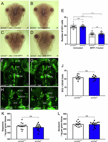
acmsd-/- zebrafish retain normal dopaminergic neuron numbers and susceptibility to MPP +. A–D In situ hybridisation against th1 showed no change in staining pattern between acmsd+/+ and acmsd-/- zebrafish either before or after MPP+ exposure. E There was no difference in th1+ neuron numbers in the ventral diencephalon between acmsd+/+ and acmsd-/- zebrafish (p = 0.9866). MPP+ exposure resulted in significant reductions in th1+ cells irrespective of genotype (43.1% in acmsd+/+; 43.4% in acmsd-/-, p < 0.0001). Total cell counts between groups following exposure remained indistinguishable (p = 0.9999). n = 26–35 per group, from three biological replicates. Statistics from a two-way ANOVA with Tukey’s multiple comparisons post-hoc test. Monoaminergic neurons were visualised using the ETvmat2:GFP transgenic line at 5dpf in the full brain (F, G dorsal view) and in the DC neuronal groups (H, I) in both acmsd+/+ and acmsd-/- zebrafish. Scale bar = 50 µm. DC diencephalic neurons, Hc caudal hypothalamus, PT pretectal neural cluster, Ra raphe nucleus, Te telencephalic neurons. J No difference in the number of neuronal cell bodies in DC2 and DC4/5 was seen between acmsd+/+ and acmsd-/- zebrafish (p = 0.8126, unpaired two-tailed t test). n = 9 per genotype, from three biological replicates.No difference in the concentrations of dopamine (K, p = 0.2808) or serotonin (L, p = 0.9079) were identified in whole 11mpf brains. Data analysed using an unpaired two-tailed t test. n = 14 acmsd-/-; 12 acmsd+/+, 1 brain per replicate.
|