
Fig. 3
- ID
- ZDB-FIG-250507-38
- Publication
- Shanbhag et al., 2025 - Pannexin-2 deficiency disrupts visual pathways and leads to ocular defects in zebrafish
- Other Figures
- All Figure Page
- Back to All Figure Page
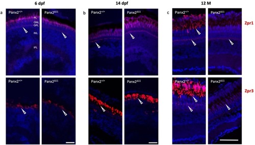
Cone and rod biomarker expression is reduced in adult Panx2 Δ11 retina. Antibody staining with Zpr1 and Zpr3 antibodies, shown in red, exhibited double cone photoreceptors, and outer segments of rods and green cones, respectively. No difference between genotypes was observed at the early stages, 6 dpf (a) and 14 dpf (b). The adult stage showed decreased Zpr1 and Zpr3 immunofluorescence in 12-month-old Panx2 Δ11 retina. Nuclei were shown in blue using DAPI. Abbreviations: photoreceptor layer (PL), outer nuclear layer (ONL), outer plexiform layer (OPL), inner nuclear layer (INL), inner plexiform layer (IPL), and 12-month-old (12 M). Scale bars: (a, b) 20 μm; (c) 50 μm. (For interpretation of the references to colour in this figure legend, the reader is referred to the web version of this article.) |
Reprinted from Biochimica et biophysica acta. Molecular basis of disease, , Shanbhag, R., Zoidl, G.S.O., Nakhuda, F., Sabour, S., Nauman, H., Zoidl, C., Bahl, A., Tabatabaei, N., Zoidl, G.R., Pannexin-2 deficiency disrupts visual pathways and leads to ocular defects in zebrafish, 167807167807, Copyright (2025) with permission from Elsevier. Full text @ BBA Molecular Basis of Disease