
Fig. 6 - Supplemental 1
- ID
- ZDB-FIG-250416-50
- Publication
- Childers et al., 2025 - Protein absorption in the zebrafish gut is regulated by interactions between lysosome rich enterocytes and the microbiome
- Other Figures
- All Figure Page
- Back to All Figure Page
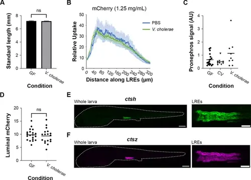
Long-term exposure to V. cholerae required to reduce protein uptake activity in lysosome-rich enterocytes (LREs) without affecting larval growth or protein availability. (A) Plot of standard length in larvae that were germ-free (GF) or monoassociated with V. cholerae. There was not a significant difference in standard length (Two-tailed t-test, p=0.66, n=18–22). (B) Plot of mCherry uptake in larvae gavaged with PBS or live V. cholerae. There was no difference in the average mCherry uptake between the conditions (two-way ANOVA, p=0.527, n=7–16). (C) Plot of mCherry fluorescence signal in pronephros of GF, conventional (CV), and V. cholerae-colonized larvae. There was no significant difference between GF, CV, and V. cholerae (one-way ANOVA, p=0.154, n=9). (D) Plot of average luminal mCherry fluorescence in GF and V. cholerae-colonized larvae at 6 dpf by 1 hr PG. Luminal mCherry fluorescence was not significantly different (Two-tailed t-test, p=0.60, n=18–20). (E-F) Confocal images of ctsh (E) and ctsz (F) hybridization chain reaction (HCR) probe localization in whole larva and the LRE region. Larva is outlined with a dashed line. Whole larva scale = 200 μm. LRE scale = 50 μm. |