
Fig. 1
- ID
- ZDB-FIG-250416-39
- Publication
- Childers et al., 2025 - Protein absorption in the zebrafish gut is regulated by interactions between lysosome rich enterocytes and the microbiome
- Other Figures
- All Figure Page
- Back to All Figure Page
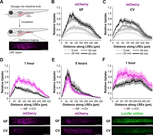
Microbes slow the rate of soluble cargo uptake in lysosome-rich enterocytes (LREs). (A) Cartoon depicting experimental design of the gavage assay in GF and CV larvae. Following derivation under conventional (CV) or germ-free (GF) conditions, 6 dpf larvae were gavaged, and uptake of luminal cargoes by LREs was measured by confocal microscopy in the LRE region (approximately 300 μm in length). (B, C) Plots of normalized mCherry fluorescence intensity along the LRE region over time in 6 dpf GF (B) and CV (C) larvae. Minutes after gavage, LREs rapidly took up and quickly accumulate mCherry in GF larvae. The anterior LREs approached full saturation by 40 min post gavage. Cargo uptake was slower in CV larvae, and anterior LREs did not reach saturation by 40 min post gavage. (D, E) Top: Plots of normalized mCherry fluorescence intensity along the LRE region of GF and CV larvae at 1 (D) and 5 (E) hr PG. GF larvae internalized significantly more mCherry than CV larvae (2-way ANOVA, p<0.0001, n=8–10) 1 hr PG, and CV larvae reached a similar level of mCherry accumulation to GF larvae by 5 hr PG (two-way ANOVA, p=0.137, n=8–11). Bottom: Representative confocal images showing mCherry signal in the LRE region (scale bars = 50 µm). (F) Top: Plot of normalized lucifer yellow fluorescence intensity along the LRE region of GF and CV larvae at 1 hr post gavage. LREs in GF larvae internalized significantly more Lucifer yellow than CV larvae by 1 hr post gavage (two-way ANOVA, p<0.0001, n=8). |