
Fig. 5
- ID
- ZDB-FIG-250415-54
- Publication
- Lin et al., 2025 - Soy Protein-Cultured Mesenchymal Stem Cell-Secreted Extracellular Vesicles Target the Neurovascular Unit: Insights from a Zebrafish Brain Injury Model
- Other Figures
- All Figure Page
- Back to All Figure Page
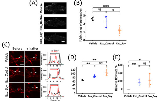
Therapeutic effect of exosomes on the function and integrity of the NVU. (A) Representative images of extravascular fluorescent tracer accumulation in the cranial region of zebrafish larvae 1 h posthypoxia, illustrating the degree of BBB disruption under each treatment condition (gray: RITC-dextran, MW: 10 k). Scale bar: 100 μm. (B) Quantitative analysis of fold change in tracer permeation, indicating BBB integrity across the Vehicle, Exo_Control, and Exo_Soy groups (n = 5 per group). (C) Left: Representative images of the cerebral vasculature in zebrafish larvae treated with Vehicle, Exo_Control, or Exo_Soy after a 15 min hypoxic insult (red: mCherry). White arrows point to the regions where vessel width measurements were taken. Right: Cross-sectional fluorescence intensity profiles along the cerebral vessels, illustrating changes before and 1 h after hypoxia in each treatment group. Scale bar: 20 μm. (D) Quantitative analysis of the relative vessel width, showing that Exo_Soy significantly preserves the vessel width compared to the Vehicle group, while Exo_Control displays a similar trend. (E) Quantitative analysis of the relative cerebral blood flow, demonstrating significant improvement in the Exo_Soy group compared to the Vehicle group, with Exo_Control showing comparable efficacy. Data are shown as mean ± SD with individual data points (n = 5 larvae per group) in panels (B), (D), and (E). |