
FIGURE
Fig. 3
- ID
- ZDB-FIG-250415-52
- Publication
- Lin et al., 2025 - Soy Protein-Cultured Mesenchymal Stem Cell-Secreted Extracellular Vesicles Target the Neurovascular Unit: Insights from a Zebrafish Brain Injury Model
- Other Figures
- All Figure Page
- Back to All Figure Page
Fig. 3
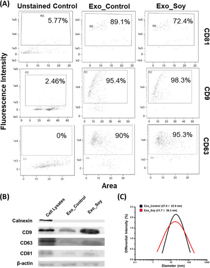
Characterization of exosomes. WJ-MSC-secreted exosomes collected from soy protein-coated (Exo_Soy) and uncoated (Exo_Control) culture dishes were, respectively, characterized for the surface markers CD81, CD9, and CD63 by (A) flow cytometric analyzer (ImageStreamX Mark II, Amnis) and (B) Western blot (Calnexin served as the negative marker of exosomes, and β-actin is the loading control). (C) DLS analysis was performed to characterize the respective diameters of these exosomes. |
Expression Data
Expression Detail
Antibody Labeling
Phenotype Data
Phenotype Detail
Acknowledgments
This image is the copyrighted work of the attributed author or publisher, and
ZFIN has permission only to display this image to its users.
Additional permissions should be obtained from the applicable author or publisher of the image.
Full text @ ACS Biomater Sci Eng