
Fig. 4
- ID
- ZDB-FIG-250415-53
- Publication
- Lin et al., 2025 - Soy Protein-Cultured Mesenchymal Stem Cell-Secreted Extracellular Vesicles Target the Neurovascular Unit: Insights from a Zebrafish Brain Injury Model
- Other Figures
- All Figure Page
- Back to All Figure Page
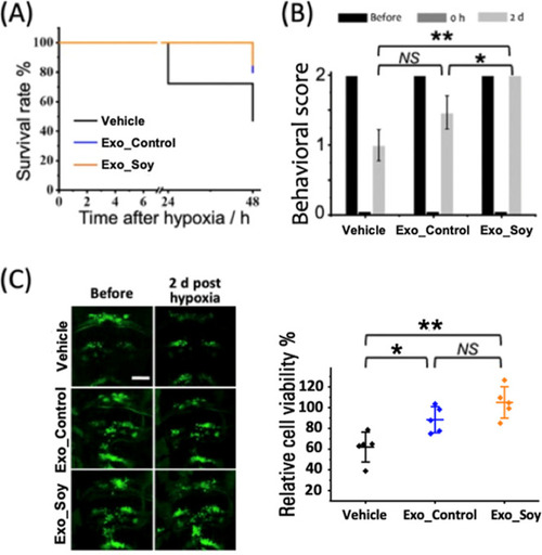
Therapeutic effect of exosomes on larval survival, neurological function, and cerebral cell viability. (A) Survival curve of zebrafish larvae (6 dpf) treated with vehicle, control exosomes (Exo_Control), or soy-derived exosomes (Exo_Soy) posthypoxia. Data represent n = 60 larvae per group. (B) Behavioral assessment of neurological function, evaluated immediately after hypoxia (0 h) and 2 days posthypoxia (2 d). Data are presented as mean ± SD from three independent experiments with n = 5 larvae per experiment. (C) Left: Representative fluorescence images of cranial motor neurons (green: GFP-labeled) in zebrafish larvae, acquired before and 2 days posthypoxia. Scale bar: 50 μm. Right: Quantification of relative cerebral cell viability at 2 days posthypoxia in the Vehicle, Exo_Control, and Exo_Soy groups. Data are presented as mean ± SD with individual data points (n = 5 per group). |