
Fig. 6
- ID
- ZDB-FIG-250415-55
- Publication
- Lin et al., 2025 - Soy Protein-Cultured Mesenchymal Stem Cell-Secreted Extracellular Vesicles Target the Neurovascular Unit: Insights from a Zebrafish Brain Injury Model
- Other Figures
- All Figure Page
- Back to All Figure Page
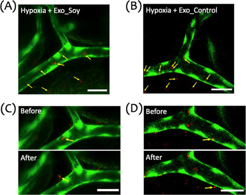
Distribution and extravasation of exosomes in a zebrafish hypoxic brain injury model. (A,B) Representative image of the cranial vasculature in zebrafish larvae subjected to a 15 min hypoxic insult, followed by microinjection of DiI-labeled Exo_Soy (A) or Exo_Control (B) exosomes. The image illustrates the distribution of exosomes within and outside the vasculature. Green represents the vasculature (GFP), and red indicates exosomes (DiI). Yellow arrows point to exosomes located both inside the vessel and those that have extravasated across the vascular wall. (C,D) Sequential images showing exosome migration and extravasation in hypoxia-exposed larvae. Panels illustrate Exo_Soy (C) and Exo_Control (D) before (top) and after (bottom) crossing the vascular wall. Yellow arrows mark exosomes transitioning from intravascular to extravascular regions. Scale bar: 20 μm. |