FIGURE

Figure 1

ID
ZDB-FIG-241212-32
Publication
Zheng et al., 2024 - Quantum Dots-caused Retinal Degeneration in Zebrafish Regulated by Ferroptosis and Mitophagy in Retinal Pigment Epithelial Cells through Inhibiting Spliceosome
Other Figures
All Figure Page
Back to All Figure Page
Figure 1

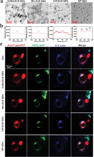
Characterizations of QDs and screening their impacts on the eye development of zebrafish. a) Representative images of QDs by TEM, scale bar, 50 nm. b) The Raman intensity of QDs. c) The lateral views of the effect of QDs (1 mg L−1) on retinal development in Tg (Atoh7:gapRFP::Ptf1a:GFP::Crx:CFPcaax) zebrafish at 168 hpf. The retinal ganglion cell layer (GCL) is labeled by red fluorescence protein (RPF), the inner nuclear layer (INL, including AC and HC) is labeled by green fluorescent protein (GFP), and cyan fluorescent protein (CFP), BC is labeled by CFP, and outer nuclear layer (ONL, composed of PR) labeled by RFP and CFP. Scale bar, 100 µm.

Expression Data

Expression Detail
Antibody Labeling
Phenotype Data

Phenotype Detail
Acknowledgments
This image is the copyrighted work of the attributed author or publisher, and ZFIN has permission only to display this image to its users. Additional permissions should be obtained from the applicable author or publisher of the image. Full text @ Adv Sci (Weinh)