
Fig. 12
- ID
- ZDB-FIG-240913-43
- Publication
- Fernezelian et al., 2024 - Mapping the cellular expression patterns of vascular endothelial growth factor aa and bb genes and their receptors in the adult zebrafish brain during constitutive and regenerative neurogenesis
- Other Figures
- All Figure Page
- Back to All Figure Page
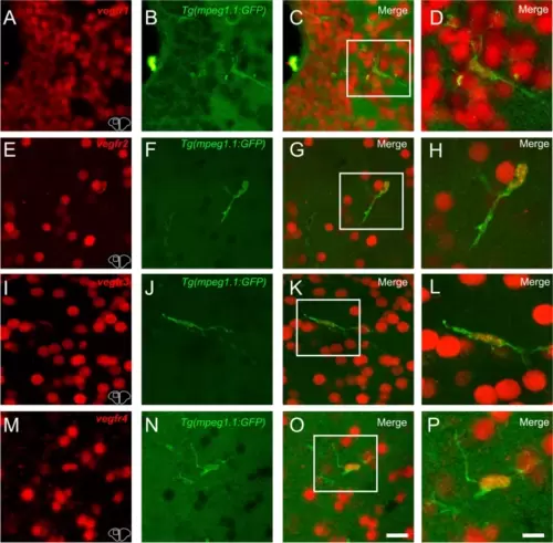
vegfr1, vegfr2, vegfr3, and vegfr4 expression in microglia/immune cells. A-C, E–G, I-K, and M–O vegfr1, vegfr2, vegfr3, and vegfr4 in situ hybridization (red) in Tg(mpeg1.1:GFP) zebrafish (green) at the level of the telencephalon. C, G, K and O Merged images showing colocalization between vegfr and microglial cells. Although expression is mostly present in other cell types, a colocalization is found between vegfr and microglia/immune cells. D, H, L and P Higher magnification view of the square in the preceded picture further presenting the vegfr expression in microglia/immune cells. Bar: 6 µm (D, H, L and P) |