
Fig. 3
- ID
- ZDB-FIG-240913-34
- Publication
- Fernezelian et al., 2024 - Mapping the cellular expression patterns of vascular endothelial growth factor aa and bb genes and their receptors in the adult zebrafish brain during constitutive and regenerative neurogenesis
- Other Figures
- All Figure Page
- Back to All Figure Page
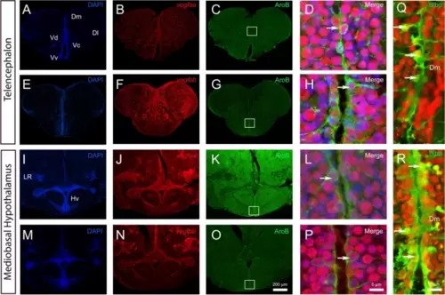
vegfaa and vegfbb are expressed in almost all neural stem cells. A-P vegfaa and vegfbb in situ hybridization (red) followed by AroB immunohistochemistry (green) in the telencephalon (A-H) and mediobasal hypothalamus (I-P) with DAPI counterstaining (blue). High magnification views of the dorsomedian telencephalon (Dm) (D and H) and of the mediobasal hypothalamus (Hv) (L and P) showing that vegfaa and vegfbb (in red) are detected in some AroB-positive neural stem cells (see arrows). White squares (C, G, K and O) highlight the respective high-power magnifications in D, H, L and P. Q and R vegfaa and vegfbb in situ hybridization (red) followed by Blbp immunohistochemistry (green) in the dorsomedian telencephalon. Arrows show examples of co-expression of vegfaa and bb with AroB and Blbp. Bars: 200 µm (A-C, E–G, I-K, M–O), 10 µm (Q and R) and 6 µm (D, H, L and P) |