
|
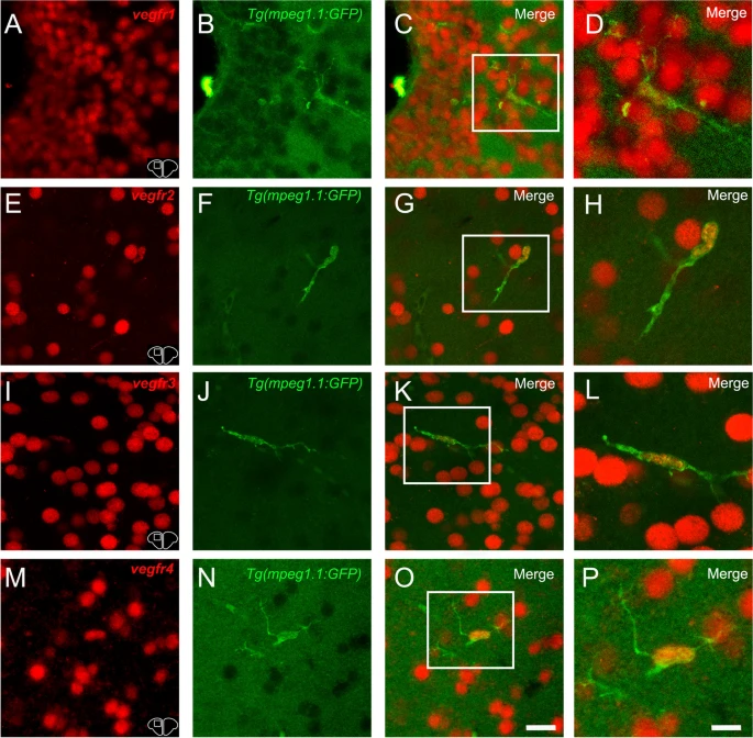
Fig. 12 vegfr1, vegfr2, vegfr3, and vegfr4 expression in microglia/immune cells. A-C, E–G, I-K, and M–O vegfr1, vegfr2, vegfr3, and vegfr4 in situ hybridization (red) in Tg(mpeg1.1:GFP) zebrafish (green) at the level of the telencephalon. C, G, K and O Merged images showing colocalization between vegfr and microglial cells. Although expression is mostly present in other cell types, a colocalization is found between vegfr and microglia/immune cells. D, H, L and P Higher magnification view of the square in the preceded picture further presenting the vegfr expression in microglia/immune cells. Bar: 6 µm (D, H, L and P)