
Fig. 2
- ID
- ZDB-FIG-240913-33
- Publication
- Fernezelian et al., 2024 - Mapping the cellular expression patterns of vascular endothelial growth factor aa and bb genes and their receptors in the adult zebrafish brain during constitutive and regenerative neurogenesis
- Other Figures
- All Figure Page
- Back to All Figure Page
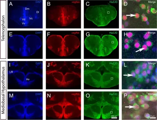
vegfaa and vegfbb are expressed in almost all neurons within telencephalon and mediobasal hypothalamus. A-P vegfaa and vegfbb in situ hybridization (red) followed by HuC/D immunohistochemistry (green) in the telencephalon (A-H) and mediobasal hypothalamus (I-P) with DAPI counterstaining (blue). D, H, L and P High magnification views of the dorsomedian telencephalon (Dm) (D and H) and of the mediobasal hypothalamus (Hv) (L and P) showing that vegfaa and vegfbb are strongly expressed in HuC/D-positive neurons (see arrows). White squares (C, G, K and O) highlight the respective high-power magnifications in D, H, L and P. Arrows show examples of co-expression of vegfaa and bb with HuC/D. Bars: 200 µm (A-C, E–G, I-K, M–O) and 6 µm (D, H, L and P) |