
Fig. 4
- ID
- ZDB-FIG-240913-35
- Publication
- Fernezelian et al., 2024 - Mapping the cellular expression patterns of vascular endothelial growth factor aa and bb genes and their receptors in the adult zebrafish brain during constitutive and regenerative neurogenesis
- Other Figures
- All Figure Page
- Back to All Figure Page
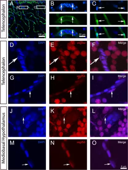
vegfaa and vegfbb are expressed in endothelial cells. A-C Representative image of Tg(fli1a:EGFP) zebrafish brain section through the telencephalon with DAPI counterstaining. B and C show high-power views of the white squares in A, with split channels showing DAPI in blue, the fli1a:EGFP transgene in green, and the merged images. Arrows indicate that elongated nuclei lining the blood vessels correspond to fli1a:EGFP-positive endothelial cells. (D-O) vegfaa and vegfbb in situ hybridization (red) with DAPI staining in the telencephalon (D-I) and mediobasal hypothalamus (J-O). vegfaa and vegfbb (in red) are expressed in some elongated nuclei localized along blood vessels and corresponding to endothelial cells (see arrows). Bar: 100 µm (A), 20 µm (B-C).6 µm (D-O) |