
Fig. 13
- ID
- ZDB-FIG-240913-44
- Publication
- Fernezelian et al., 2024 - Mapping the cellular expression patterns of vascular endothelial growth factor aa and bb genes and their receptors in the adult zebrafish brain during constitutive and regenerative neurogenesis
- Other Figures
- All Figure Page
- Back to All Figure Page
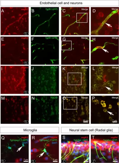
Vegfr3 and Vegfr4 are expressed in neurons, endothelial cells and neural stem cells. A-T Vegfr3 and Vegfr4 immunostainings (red) in adult zebrafish telencephalon. A-D and E–H Vegfr3 (A-D) and Vegfr4 (E–H) expression (red) in endothelial cells (green) from Tg(fli1a:EGFP) zebrafish (see arrows). I-L and M-P Vegfr3 (I-L) and Vegfr4 (M-P) expression (red) in HuC/D -positive cells (neurons) cells in adult zebrafish (see arrows). D, H, L and P High magnifications views of the respective white square. Q, R Merged images showing no colocalization between Vegfr3 (Q) and Vegfr4 (R) in microglial cells/immune cells (green) from Tg(mpeg1.1:GFP) (see arrows). (S-T) Merged images showing colocalization between Vegfr3 (Q) and Vegfr4 (R) in neural stem cells (green) from Tg(GFAP::GFP) (see arrows). Bars: 35 µm (A-C, E–G, I-K) Bar: 7 µm (D, H, L, P, Q, R, S and T) |