
Fig. 11
- ID
- ZDB-FIG-240913-42
- Publication
- Fernezelian et al., 2024 - Mapping the cellular expression patterns of vascular endothelial growth factor aa and bb genes and their receptors in the adult zebrafish brain during constitutive and regenerative neurogenesis
- Other Figures
- All Figure Page
- Back to All Figure Page
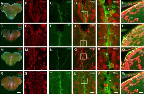
vegfr genes are expressed in neural stem cells. A-R vegfr1, vegfr2, vegfr3, and vegfr4 in situ hybridization (red) followed by AroB immunohistochemistry (green) in the telencephalon with DAPI counterstaining (blue). B, H, M, and S High magnification views of the ventricular layer at the levels of the red squares in the telencephalon showing vegfr gene expression along the ventricular layer. C, I, N and T High magnification views of the respective red squares showing AroB-positive cells. D, J, O and U Merged pictures showing the colocalization between the vegfr and the AroB-positive cells. The asterisks (*) and arrows (last columns) represent the absence or presence of vegfr expression in neural stem cells, respectively. F, L, Q and W vegfr1, vegfr2, vegfr3, and vegfr4 in situ hybridization (red) followed by Blbp immunohistochemistry (green) in the dorsomedian telencephalon (Dm) with DAPI counterstaining (blue) showing that almost all Blbp-positive neural stem cells are vegfr-positive (see arrows). Bar: 200 µm (A, G, M, and R), 30 µm (B-D, H-J, M–O and S-U), 15 µm (F, L, Q, and W), 7 µm (E, K, P, and V) |