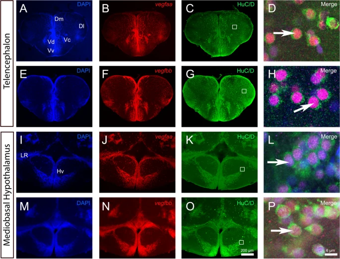
Fig. 2 vegfaa and vegfbb are expressed in almost all neurons within telencephalon and mediobasal hypothalamus. A-P vegfaa and vegfbb in situ hybridization (red) followed by HuC/D immunohistochemistry (green) in the telencephalon (A-H) and mediobasal hypothalamus (I-P) with DAPI counterstaining (blue). D, H, L and P High magnification views of the dorsomedian telencephalon (Dm) (D and H) and of the mediobasal hypothalamus (Hv) (L and P) showing that vegfaa and vegfbb are strongly expressed in HuC/D-positive neurons (see arrows). White squares (C, G, K and O) highlight the respective high-power magnifications in D, H, L and P. Arrows show examples of co-expression of vegfaa and bb with HuC/D. Bars: 200 µm (A-C, E–G, I-K, M–O) and 6 µm (D, H, L and P)
                
                    
                        Image


                        Figure Caption


                        Acknowledgments


This image is the copyrighted work of the attributed author or publisher, and
ZFIN has permission only to display this image to its users.
Additional permissions should be obtained from the applicable author or publisher of the image.
      Full text @ Neural Dev.