
Fig. 4
- ID
- ZDB-FIG-240802-45
- Publication
- Kumar et al., 2024 - Whole-body replacement of larval myofibers generates permanent adult myofibers in zebrafish
- Other Figures
- All Figure Page
- Back to All Figure Page
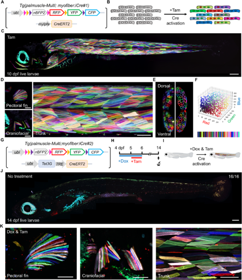
Multicolor barcoding of the entire myofiber population in palmuscle-Multi zebrafish. (A) The palmuscle-Multi and myofiber:iCre#1 transgenic constructs. (B) Schematic drawing of individual myofibers before and after Cre activation. Addition of tamoxifen (Tam) activates Cre recombinase, which acts on Brainbow-based cassettes to convert label-free myofibers into color-barcoded myofibers. (C) Whole-animal view of a live palmuscle-Multi zebrafish larva at 10 dpf. (D) Magnified view of the pectoral fin, craniofacial, and trunk myofibers. (E) Representative cross-sectional view of a myotome (left). Schematic outlines of color-barcoded myofibers are shown on the right. (F) Color space analysis of 2057 individual myofibers from 32 myotomes captured from a single palmuscle-Multi. About 50 distinct hues were detected in live animals upon Cre activation. (G) The palmuscle-Multi and myofiber:iCre#2 transgenic constructs. (H) Timeline of the treatment and tracking scheme. (I) Schematic drawing of the Tg(palmuscle-Multi; myofiber:iCre#2) larva before and after Cre activation. (J) Whole-animal view of the Tg(palmuscle-Multi; myofiber:iCre#2) larva without Dox and Tam treatment. (K) The pectoral fin, craniofacial and trunk region displayed multicolor myofibers upon treatment with Dox and Tam. n = number of animals (J). Stitched image (C, D, J). Scale bars, 200 µm (C, J); 100 µm (D, K). dpf, days post-fertilization. Source data are available online for this figure. |