
Fig. 5
- ID
- ZDB-FIG-240703-112
- Publication
- He et al., 2024 - Meningeal lymphatic supporting cells govern the formation and maintenance of zebrafish mural lymphatic endothelial cells
- Other Figures
- All Figure Page
- Back to All Figure Page
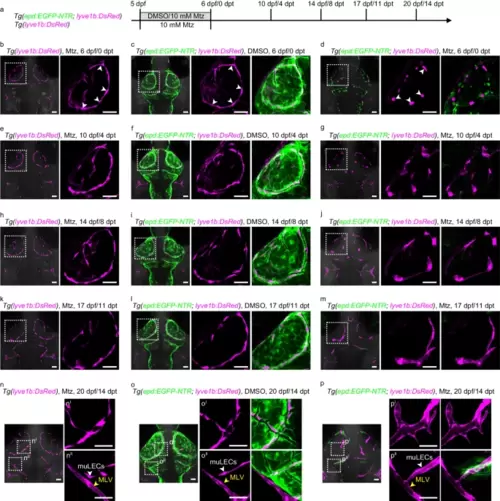
The mLSCs are essential for maintaining the dispersed distribution pattern of muLECs at the larval stage.a Schematic diagram showing experimental design for meningeal lymphatic supporting cell (mLSC) ablation. b, c, e, f, h, i, k, l, n, o Dorsal confocal images of mural lymphatic endothelial cells (muLECs) and mLSCs in brains that did not induce muLEC or mLSC injury at 6 dpf/0 dpt, 10 dpf/4 dpt, 14 dpf/8 dpt, 17 dpf/11 dpt and 20 dpf/14 dpt. White arrowheads indicate muLECs. Yellow arrowheads indicate meningeal lymphatic vessels (MLV). n = 20 per experiment. d, g, j, m, p Dorsal confocal images showing the progressive formation of lymphatic vessels from collapsed muLECs after inducing injury to mLSCs. White arrowheads in (d) indicate collapsed muLECs. White arrowheads in (p) indicate muLEC-derived lymphatic vessels. Yellow arrowheads indicate MLV. d n = 28; g n = 24; j n = 20; m n = 17; p n = 19. The experiments were repeated three times independently with similar results. The white dashed boxes outline the enlarged areas. Scale bars: 50 µm. |