Fig. 4
- ID
- ZDB-FIG-240620-47
- Publication
- Gauthier et al., 2024 - Therapeutic antibody engineering for efficient targeted degradation of membrane proteins in lysosomes
- Other Figures
- All Figure Page
- Back to All Figure Page
|
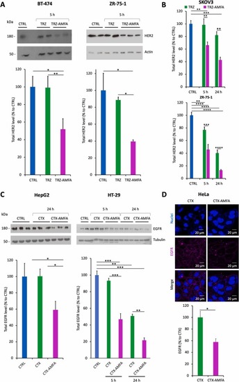
Degradation of HER2 and EGFR proteins. (A) HER2 degradation analysis in BT-474 and ZR-75–1 cells at 5 h by Western blot. Cells were treated with 5 nM TRZ or TRZ-AMFA for 5 h (n=2) and analyzed by Western blot. (B) HER2 degradation at 5 h and 24 h in SKOV3 and ZR-75–1 cells. Cells were treated with 5 nM TRZ or TRZ-AMFA for 5 h or 24 h and HER2 levels were determined by ELISA (n=2). (C) EGFR degradation in HepG2 (left) or HT-29 cells (right). Cells were treated with 10 nM CTX or CTX-AMFA for 5 h or 24 h. One representative experiment out of two is shown. For Fig. A-C, membrane receptor levels were quantified and expressed as the percentage ± SEM of total HER2 or EGFR levels in control cells, accordingly. (D) EGFR membrane detection by confocal microscopy. HeLa cells were treated with 10 nM CTX or CTX-AMFA for 24 h and EGFR was detected with an anti-EGFR antibody together with a secondary antibody coupled with AlexaFluor647®. Microscopy was performed on living cells and EGFR quantification is presented as the mean fluorescence ± SEM of antibody per cell determined in 40 cells. Statistical analysis was performed with Tukey’s multiple comparison test and Student’s t-test:* p value < 0.05; ** p value < 0.01; *** p value < 0.001; **** p value < 0.0001. |

