Fig. 3
- ID
- ZDB-FIG-240620-46
- Publication
- Gauthier et al., 2024 - Therapeutic antibody engineering for efficient targeted degradation of membrane proteins in lysosomes
- Other Figures
- All Figure Page
- Back to All Figure Page
|
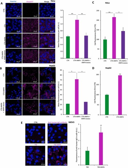
Internalization of mAb and mAb-AMFA into cells. (A-B) Internalization of CTX or CTX-AMFA by confocal microscopy. HeLa cells (A) or HepG2 cells (B) were treated for 5 h with 5 nM CTX or CTX-AMFA which were detected with 2.5 nM anti-human IgG antibody coupled to AlexaFluor647Ⓡ. A·20 mM AMFA excess was used to prevent the cell uptake mediated by M6PR. Results are presented as the mean fluorescence ± SEM per cell determined in 30 cells. (C-D). Internalization of CTX and CTX-AMFA by flow cytometry. HeLa (C) and HepG2 cells (D) were treated with 5 nM CTX or CTX-AMFA labeled with AlexaFluor488Ⓡ for 5 h. An excess of 20 mM AMFA was used to inhibit CTX-AMFA cell uptake mediated by M6PR. Data are expressed as the percentage ± SEM of cell fluorescence compared to CTX (n=2). (E) Internalization of TRZ and TRZ-AMFA by confocal microscopy. SKOV3 cells were treated with 5 nM TRZ or TRZ-AMFA and with 2.5 nM anti-human IgG antibody coupled with AlexaFluor647Ⓡ for 24 h. Microscopy was performed on living cells and data are expressed as the mean fluorescence ± SD of antibody per cell quantified in 60 cells. Tukey’s test was performed for multiple comparisons. Student’s t-test was performed for paired analysis: * p value < 0.05; ** p value < 0.01. |

