
Fig. 5
- ID
- ZDB-FIG-240522-5
- Publication
- Akçaöz-Alasar et al., 2024 - Epitranscriptomics m6 A analyses reveal distinct m6 A marks under tumor necrosis factor α (TNF-α)-induced apoptotic conditions in HeLa cells
- Other Figures
- All Figure Page
- Back to All Figure Page
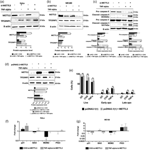
Western blot analyses of METTL3 knockdown cells treated with TNF-α. Cells were transfected with 25 nM control si-NC or si-METTL3 for 72 h and incubated with 2.5 ug/mL CHX or 37.5 ng/mL TNF-α for 24 h. Western blot assays were performed to examine TP53INP1 expression in METTL3 knockdown and/or TNF-α-treated HeLa (a) and ME180 (b) cells. Total cellular extracts were also assayed for caspase−3 and 8 amounts (c). Western blot assays (d) and apoptotic measurement (e) were performed with crude extracts obtained from HeLa cells transfected with the control pcDNA3.1(+) or pcDNA3.1(+) + METTL3 plasmids and treated with 37.5 ng/mL TNF-α. qPCR analyses of TP53INP1 target genes (P21, PIG3, BAX, and MDM2) in CHX treatment with METTL3 knockdown and TNF-α treatment with METTL3 knockdown HeLa (f) and ME180 (g) cells. For all western blot experiments, equal amounts of total proteins (25 μg/lane) were fractionated through a 10% SDS-PAGE. Band intensities were normalized against β-actin used as a loading control. n = 3. Two-tailed Student's t test was performed to determine the statistical significance among groups. Data presented as mean ± SD. *p ≤ 0.05, **p ≤ 0.01. |