|
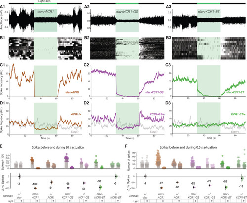
Spontaneous spiking in a larval abdominal nerve is silenced by KCR1. A Representative traces from extracellular recordings of Drosophila larvae abdominal nerve 3. ACR1, KCR1-GS, and KCR1-ET channelrhodopsins were expressed pan-neuronally with elav-Gal4. Actuation with green light (40 μW/mm2) was induced for 30 s (schematic on top). For each genotype n = 1 biologically independent sample over 1 independent experiment. B Rasters of action-potential occurrences in larvae expressing elav > ACR1, elav > KCR1-GS, and elav > KCR1-ET. For each genotype, n = 3 biologically independent samples over 3 independent experiments. C Averaged spike-frequency charts for the test genotypes. D A reduction in action potentials was detected in ACR1/+ and KCR1-GS/+ controls, but not in KCR1-ET/+ or in elav/+ controls. For each genotype in C, D, n = 3 biologically independent samples over 3 independent experiments. Error bars show 95% CI. E Quantification of spike totals in the 30 s epochs before and during actuation. Each dot in the scatter plot (top) represents the number of spikes per recording. The differences in spike counts before versus during actuation are shown (bottom). F Quantification of spike totals using 0.5 s light actuation. Error bars in E and F show 95% CI. For each genotype, n = 3 biologically independent samples over 3 independent experiments. Additional information on effect-size statistics is presented in Supplementary Dataset 1. Source data are provided as a Source Data file.
|