FIGURE
Fig. 2
- ID
- ZDB-FIG-240130-18
- Publication
- Mi et al., 2023 - Stimulation of liver fibrosis by N2 neutrophils in Wilson's disease
- Other Figures
- All Figure Page
- Back to All Figure Page
Fig. 2
|
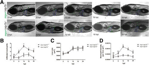
Neutrophils infiltrate into livers in atp7b-/- zebrafish indicated by mpx-positive neutrophils. (A) Representative images of mpx+/atp7b+/+ and mpx+/atp7b-/- fish after 0, 6, 12, 18, and 24 hours of Cu treatment. The livers are outlined in blue dashed lines. hpt, hours post-treatment. Scale bar: 200 μm. (B) Neutrophil count, (C) liver size, and (D) neutrophil density in wild-type and mutant fish livers. Data are the means ± SEM, n = 10 fish /group. ∗P < .05, ∗∗P < .01. |
Expression Data
Expression Detail
Antibody Labeling
Phenotype Data
Phenotype Detail
Acknowledgments
This image is the copyrighted work of the attributed author or publisher, and
ZFIN has permission only to display this image to its users.
Additional permissions should be obtained from the applicable author or publisher of the image.
Full text @ Cell Mol Gastroenterol Hepatol

