Fig. 16
- ID
- ZDB-FIG-240130-32
- Publication
- Mi et al., 2023 - Stimulation of liver fibrosis by N2 neutrophils in Wilson's disease
- Other Figures
- All Figure Page
- Back to All Figure Page
|
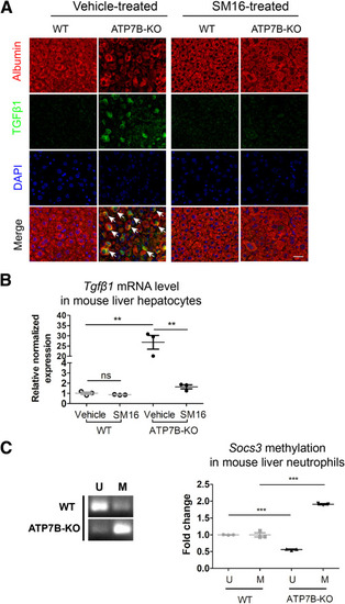
TGFβ1 is highly increased in hepatocytes in ATP7B-KO mice. (A) Formalin-fixed mouse liver tissue sections from SM16-treated or vehicle-treated wild-type and ATP7B-KO mice were subjected to immunofluorescence staining of anti-TGFβ1 and anti-albumin. Representative images of TGFβ1 (green), albumin (red), and nuclei (blue) are shown. Arrows indicate TGFβ1-positive hepatocytes. Scale bar: 40 μm. (B) qPCR of mRNA levels of Tgfβ1 in liver hepatocytes isolated from wild-type and ATP7B-KO mice upon SM16 or vehicle treatment. n = 3 sets of liver hepatocytes pooled from 2∼3 mice. ∗∗P < .01. (C) Methylation-specific PCR in Socs3 promoter in liver neutrophils from wild-type and ATP7B-KO mice. Left panel: Representative images of methylation-specific PCR in Socs3 promoter. Right panel: Quantification of band levels by ImageJ. M, methylated; U, unmethylated. n = 3 sets of liver neutrophils pooled from 2∼3 mice. ∗∗∗P < .001. DAPI, 4′,6-diamidino-2-phenylindole; WT, wild-type. |

