FIGURE

Figure 6

ID
ZDB-FIG-240113-15
Publication
Madera et al., 2023 - Gene Characterization of Nocturnin Paralogues in Goldfish: Full Coding Sequences, Structure, Phylogeny and Tissue Expression
Other Figures
All Figure Page
Back to All Figure Page
Figure 6

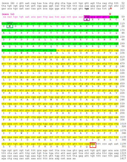
Nucleotide and deduced amino acid sequences of goldfish noc-bb. Both sequences are accessible through GenBank. Splice Variant III is composed of exon 1c in purple (coding sequence with purple background), exon 2 (green background), and exon 3 (yellow background) (accession no. OR651299). Splice Variant II is composed of non-coding exon 1b, exon 2, and exon 3 (accession no. OR651300). Red arrow indicates the insertion point of exon 1b or exon 1c on exon 2. Bold font indicates stop codons upstream of initial methionine codon. Coding region extends from the first methionine residue (purple box for variant III, green box for variant II) to the stop codon (* and red box). Backgrounds colored purple, green and yellow indicate exons 1c, 2, and 3, respectively.

Expression Data

Expression Detail
Antibody Labeling
Phenotype Data

Phenotype Detail
Acknowledgments
This image is the copyrighted work of the attributed author or publisher, and ZFIN has permission only to display this image to its users. Additional permissions should be obtained from the applicable author or publisher of the image. Full text @ Int. J. Mol. Sci.