FIGURE

Figure 7

ID
ZDB-FIG-240113-16
Publication
Madera et al., 2023 - Gene Characterization of Nocturnin Paralogues in Goldfish: Full Coding Sequences, Structure, Phylogeny and Tissue Expression
Other Figures
All Figure Page
Back to All Figure Page
Figure 7

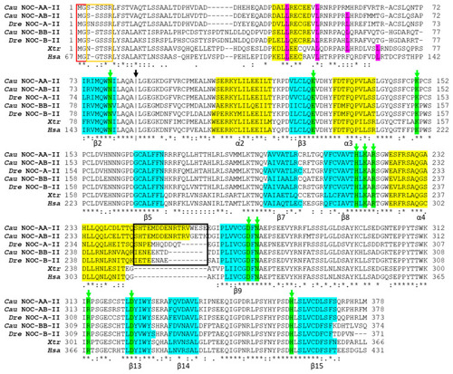
Alignment of the deduced amino acid sequences of splicing variant II of goldfish NOC-AA (Cau NOC-AA-II: WNX29031), NOC-AB (Cau NOC-AB-II: WNX29026), and NOC-BB (Cau NOC-BB-II: WNX29028) with NOC sequences from Danio rerio (Dre NOC-A-II: XP_005169676.1) and (Dre NOC-B-II: XP_021331689.1), Xenopus tropicalis (Xtr: KAE8630336.1), and Homo sapiens (Hsa: NP_036250.2). Multiple sequence alignment was conducted using Clustal v.X2. Bottom symbols (. : *), indicating a significant conservation from minor to major. Horizontal hyphens indicate gaps introduced to optimize the alignment. The vertical hyphen line (black arrow) marks the transition between exon 2 and 3. The secondary structure was predicted using JPred4. Structural elements are indicated as follows: yellow boxes (α-helix) and blue boxes (β-sheet). The names of α and β follow the nomenclature of Abshire and coworkers [27]. Green boxes pointed to by a green arrow indicate the amino acids conserved in the active site. Pink boxes mark the position of the heptad leucine repeat. Red rectangles indicate putative myristoylation sites, and the orange rectangle indicates a Ser/Thr-rich region. Black rectangles indicate the non-conserved domain of goldfish NOC paralogues compared to human and frog NOC.

Expression Data

Expression Detail
Antibody Labeling
Phenotype Data

Phenotype Detail
Acknowledgments
This image is the copyrighted work of the attributed author or publisher, and ZFIN has permission only to display this image to its users. Additional permissions should be obtained from the applicable author or publisher of the image. Full text @ Int. J. Mol. Sci.