FIGURE

Figure 13

ID
ZDB-FIG-240113-23
Publication
Madera et al., 2023 - Gene Characterization of Nocturnin Paralogues in Goldfish: Full Coding Sequences, Structure, Phylogeny and Tissue Expression
Other Figures
All Figure Page
Back to All Figure Page
Figure 13

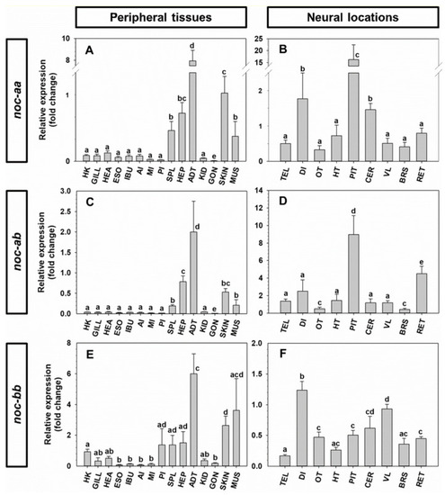
Expression of noc-aa, noc-ab and noc-bb in peripheral (A,C,E) and neural tissues of goldfish. Data are expressed as mean + S.E.M. (n = 6) relative to the hepatopancreas (A,C,E) and to the diencephalon (B,D,F). Different letters indicate statistical differences among tissues (ANOVA-SNK; p < 0.05). Head kidney (HK), gill (GILL), heart (HEA), esophagus (ESO), intestinal bulb (IBU), anterior intestine (AI), middle intestine (MI), posterior intestine (PI), spleen (SPL), hepatopancreas (HEP), adipose tissue (ADT), caudal kidney (KID), gonad (GON), skin (SKIN), muscle (MUS), telencephalon (TEL), diencephalon (DI), optic tectum (OT), hypothalamus (HT), pituitary (PIT), cerebellum (CER), vagal lobe (VL), brainstem (BRS), retina (RET).

Expression Data

Expression Detail
Antibody Labeling
Phenotype Data

Phenotype Detail
Acknowledgments
This image is the copyrighted work of the attributed author or publisher, and ZFIN has permission only to display this image to its users. Additional permissions should be obtained from the applicable author or publisher of the image. Full text @ Int. J. Mol. Sci.