
Fig. 5
- ID
- ZDB-FIG-220831-20
- Publication
- Picton et al., 2022 - Developmental switch in the function of inhibitory commissural V0d interneurons in zebrafish
- Other Figures
- All Figure Page
- Back to All Figure Page
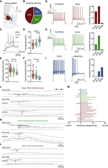
Figure 5. Intrinsic properties and morphologies of V0d interneuron subtypes in juvenile zebrafish (A) Plot of the soma position of recorded V0d interneurons showing no topographical organization of V0d interneuron subtypes. (B) Proportion of each V0d interneuron subtype identified among recorded neurons showing that the majority were slow or intermediate. (C) Recording showing a single action potential of slow, intermediate, and fast V0d interneurons. (D) Spike threshold was lowest for slow and highest for fast V0d interneurons (n = 52 slow V0d interneurons; n = 37 intermediate V0d interneurons; n = 26 fast V0d; ∗∗∗∗p < 0.0001; repeated-measures one-way ANOVA). (E) Resting membrane potential was most depolarized for slow and hyperpolarized for fast V0d interneurons (n = 52 slow V0d interneurons; n = 37 intermediate V0d interneurons; n = 26 fast V0d; ∗∗p < 0.01; ∗∗∗∗p < 0.0001; repeated-measures one-way ANOVA). (F) Input resistance was highest for slow and lowest for fast V0d interneurons (n = 52 slow V0d interneurons; n = 37 intermediate V0d interneurons; n = 26 fast V0d; ∗p < 0.05; ∗∗∗∗p < 0.0001; repeated-measures one-way ANOVA). (G) Left: example of a bursting and tonic slow V0d interneuron. Right: the proportion of each firing type among slow V0d interneurons. (H) Left: example of a bursting and tonic intermediate V0d interneuron. Right: the proportion of each firing type among intermediate V0d interneurons. (I) Left: example of a tonic and adapting fast V0d interneuron. Right: the proportion of each firing type among fast V0d interneurons. (J) Morphological reconstructions of neurobiotin-filled slow V0d interneurons. The top example shows a side view, and the bottom two examples show a top-down view to illustrate the commissural axon projection. The dashed line indicates the midline of the spinal cord. (K) Morphological reconstructions of neurobiotin-filled intermediate V0d interneurons. (L) Morphological reconstructions of neurobiotin-filled fast V0d interneurons. (M) Plot of the commissural axon projection direction and distances of the identified slow, intermediate, and fast V0d interneurons. |