
Fig. 3
- ID
- ZDB-FIG-220831-18
- Publication
- Picton et al., 2022 - Developmental switch in the function of inhibitory commissural V0d interneurons in zebrafish
- Other Figures
- All Figure Page
- Back to All Figure Page
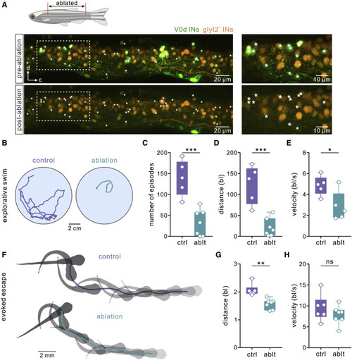
Figure 3. Ablation of V0d interneurons in juvenile zebrafish affects spontaneous swimming (A) Confocal images of the spinal cord of a juvenile zebrafish (6 wpf) showing V0d interneurons before (“pre-ablation”) and after two-photon laser ablation (“post-ablation”). The panel on the right shows the expansion of the area indicated by a dashed line. V0d interneurons are shown in green and non-V0d glycinergic interneurons in orange. (B) Tracking of in vivo spontaneous swimming in control and following the ablation of V0d interneurons in juvenile zebrafish. (C) Ablation of V0d interneurons significantly reduced the number of spontaneous swim episodes (n = 5 control animals; n = 7 ablated animals; ∗∗∗p < 0.001; two-tailed Student’s t test). (D) Ablation of V0d interneurons significantly reduced the overall spontaneous swim distance (n = 5 control animals; n = 7 ablated animals; ∗∗∗p < 0.001; two-tailed Student’s t test). (E) Ablation of V0d interneurons significantly reduced the overall swim velocity (n = 5 control animals; n = 7 ablated animals; ∗p < 0.05; two-tailed Student’s t test). (F) A sequence of escape and fast swimming evoked by a sound stimulus in control and in a V0d-ablated juvenile zebrafish. (G) Ablation of V0d interneurons significantly reduced the overall escape distance (n = 6 control animals; n = 7 ablated animals; ∗∗p < 0.01; Mann-Whitney test). (H) Ablation of V0d interneurons had no significant effect on the velocity of the escape sequence (n = 6 control animals; n = 7 ablated animals; two-tailed Student’s t test). See also Figure S1. |