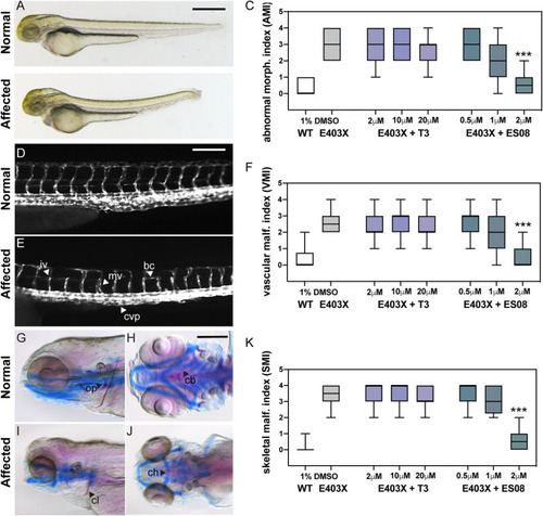
Effect of T3 and ES08 on E403X mutant TRα-expressing zebrafish embryos and E403X TRα mutation-containing, patient-derived inducible pluripotent stem cells. (A and B) Normal or abnormal morphology of embryos injected with hTRα WT or hTRα E403X mRNAs at 3 days postfertilization (dpf). Cerebral edema (ce), pericardial edema (pe), altered body curvature (bc), and thickening of caudal vein plexus (cvp) were used to calculate an abnormal morphology index (AMI, 0 = unaffected, 1 to 4 = affected). Scale bar = 250 μm. Boxplots of AMI for WT (open) or E403X injected zygotes treated with DMSO (gray), T3 (purple), or ES08 (gray-green) are shown in panel C. (D and E) transgenic tg(kdrl:EGFP) line was used to visualize normal or disrupted vascular architecture of the trunk-tail region of embryos at 4 dpf (lateral view, head to the left). Anomalies, including incomplete vessel (iv), aberrant branching (ab), misplaced vessel (mv), and reduced caudal vein plexus (cvp) area, shown in panel E, were used to calculate a vascular malformation index (VMI 0 = unaffected, 1 to 4 = affected). Scale bar = 50 μm. Boxplots of VMI of WT and E403X injected zygotes treated with DMSO (gray), T3 (purple), or ES08 (gray-green) are shown in panel F. (G to J) Normal or defective cranial cartilage development and bone mineralization in 5 dpf embryos stained with Alcian Blue (in blue) and Alizarine (in red), respectively. Absent or reduced mineralization of cleitrum (cl) and operculum (op) (visible in lateral view), and malformations of ceratohyal (ch) and the five ceratobranchial (cb) arches of cartilages (visible in ventral view) were quantified to compute a skeletal malformation index (SMI). Scale bar = 100 μm. (K) Box plots of SMI of WT and E403X mutant TRα-expressing embryos following exposure to DMSO, T3, or ES08. Indices shown are mean ± SEM for pools of 60 embryos per condition, from at least 4 independent experiments. P < 0.001 (***) for comparison of malformation indices in E403X TRα-expressing embryos after exposure to DMSO versus exposure to ES08.
|
