
FIG 4
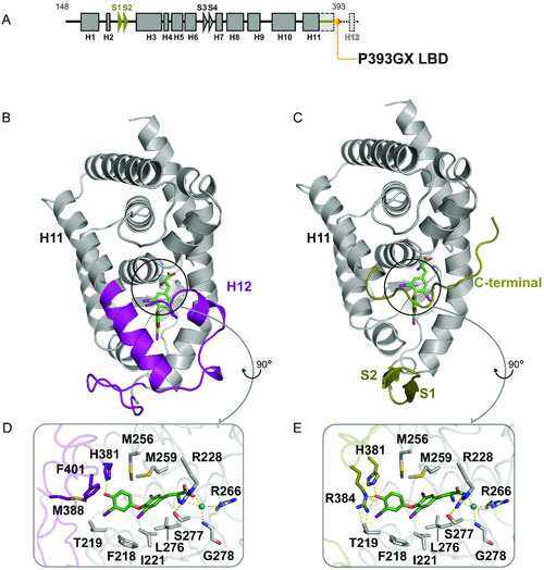
Comparison of wild-type and P393GX LBD structures. (A) Schematic representation of the P393GX LBD with alpha-helices (squares), beta-strands (arrowheads), and specific features of the P393GX mutant compared to the wild-type (highlighted in yellow). (B and C) Wild-type (B) and P393GX (C) LBD structures showing significantly different regions colored differently, with wild-type (purple) and P393GX (yellow). (D and E) Comparison of the wild-type (D) and P393GX (E) ligand-binding pockets, showing the different orientation of His 381 and different positions and interactions of Arg 384 residues. Key residues and helices are labeled with dashed lines denoting polar contacts. |