
Figure 3
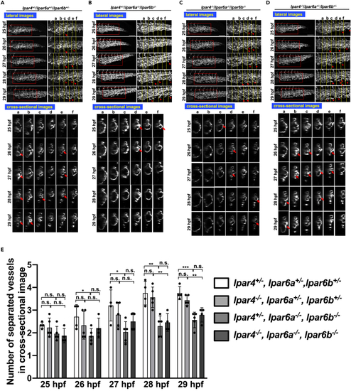
Similar abnormal CVP structure in lpar6a/lpar6b double mutant embryos (A–D) Sequential time-lapse images of control (lpar4+/−/lpar6a+/−/lpar6b+/−) (A) and lpar4+/−/lpar6a−/−/lpar6b−/− (B), lpar4−/−/lpar6a−/−/lpar6b−/− (C), and lpar4−/−/lpar6a+/−/lpar6b+/− (D) embryos at the indicated time points. Both projection views from the lateral side (upper panel) and sectional images (lower panel) are shown. Enlarged images of the area surrounded by squares are positioned in the right side (upper panel). In all embryos, endothelial cell (EC) sprouts and their anastomosis were observed although less frequent in lpar4+/−/lpar6a−/−/lpar6b−/− (B) and lpar4−/−/lpar6a−/−/lpar6b−/− (C) embryos. Arrowheads and hollow arrowheads indicate sprouts and anastomosed (re-joined) sprouts, respectively. Cross-linked structure formed in lumen is pointed with arrows. Vessel subdivision was significantly attenuated in lpar4+/−/lpar6a−/−/lpar6b−/− (B) and lpar4−/−/lpar6a−/−/lpar6b−/− embryos (C), which resulted in the remaining large lumens, as was observed for atxb−/− mutants. Note that both EC sprouting and the sign of forming the bridging structure are still observed, even less frequently. Scale bars, 100 μm. (E) Numbers of separated vessels surrounded by endothelial cells in a cross section (somite a-f in Figures 3A–3D) at the indicated timepoints. Four or five embryos with genotypes shown were evaluated (lpar4+/−lpar6a+/−lpar6b+/− n = 4, lpar4−/−lpar6a+/−lpar6b+/− n = 5, lpar4+/−lpar6a−/−lpar6b−/− n = 5, lpar4−/−lpar6a−/−lpar6b−/− n = 4). All data were expressed as means and SD. p value was calculated by the student’s t test (∗p < 0.05; ∗∗p < 0.01; ∗∗∗p < 0.001; n.s., no significance). See also Figure S7. |