
Fig. 8
- ID
- ZDB-FIG-211006-8
- Publication
- Leach et al., 2021 - The immune response is a critical regulator of zebrafish retinal pigment epithelium regeneration
- Other Figures
- All Figure Page
- Back to All Figure Page
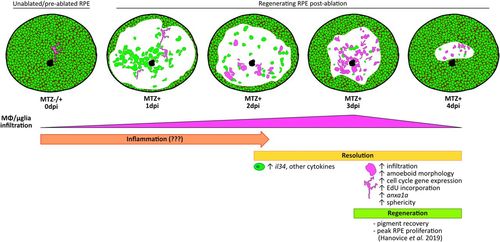
Phases of immune involvement during RPE regeneration. Schematic showing few ramified MΦs/μglia (magenta) present in the RPE (green) of unablated larvae. Infiltration of MΦs/μglia to the central RPE injury site after ablation begins at 2 dpi, peaks at 3 dpi, and wanes by 4 dpi, representing a time window when inflammation is likely resolved (2 to 4 dpi; yellow). During resolution, MΦs/μglia appear amoeboid in morphology, proliferate and express phagocytosis markers (e.g., anxa1a), and RPE express il34 and other cytokines. Peak RPE layer proliferation and recovery of pigment occurs between 3 to 4 dpi (18). This coupled with the decreased presence of MΦs/μglia in the RPE by 4 dpi may hint to a time window after ablation (3 to 4 dpi; green) when inflammation has been resolved, enabling peak RPE regeneration. |