
Fig. 2
- ID
- ZDB-FIG-211006-2
- Publication
- Leach et al., 2021 - The immune response is a critical regulator of zebrafish retinal pigment epithelium regeneration
- Other Figures
- All Figure Page
- Back to All Figure Page
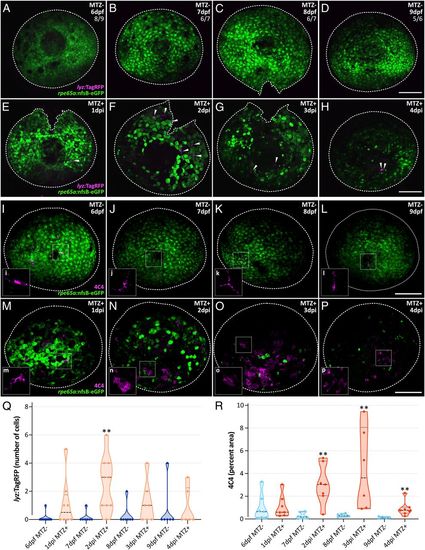
Leukocyte infiltration into the RPE injury site during regeneration. (A–H) Confocal micrographs of MTZ− (A–D) and MTZ+ (E–H) Tg(lyz:TagRFP;rpe65a:nfsB-eGFP) whole-mount eyes. Ratios at top right (A–D) indicate number of eyes lacking lyz:TagRFP+ neutrophils (white arrowheads in E–H over total number of eyes). (I–P) Fluorescent confocal micrographs of MTZ− (I–L) and MTZ+ (M–P) Tg(rpe65a:nfsB-eGFP) whole-mount eyes labeled with 4C4 to mark MΦs/μglia. (I–P) Insets show digital zooms of single cells or cell clusters to highlight 4C4+ cell morphologies. (A–P) White dashed lines designate edges of eyes. Magenta labels endogenous TagRFP or 4C4 and green labels endogenous eGFP. (Scale bars, 100 μm.) (Q) Violin plots showing a significant increase in the number of lyz:TagRFP+ neutrophils at 2 dpi when compared with 7 dpf controls. A maximum of six infiltrating cells were present at 2 dpi (datapoint in F). (R) Violin plots showing significant increases in 4C4+ staining from 2 to 4 dpi (MTZ+) when compared with MTZ− controls. (Q and R) Dashed black lines represent the median, and dotted black lines represent quartiles. SI Appendix, Table S12 contains statistical information. Dorsal is up; **P value ≤0.01. |