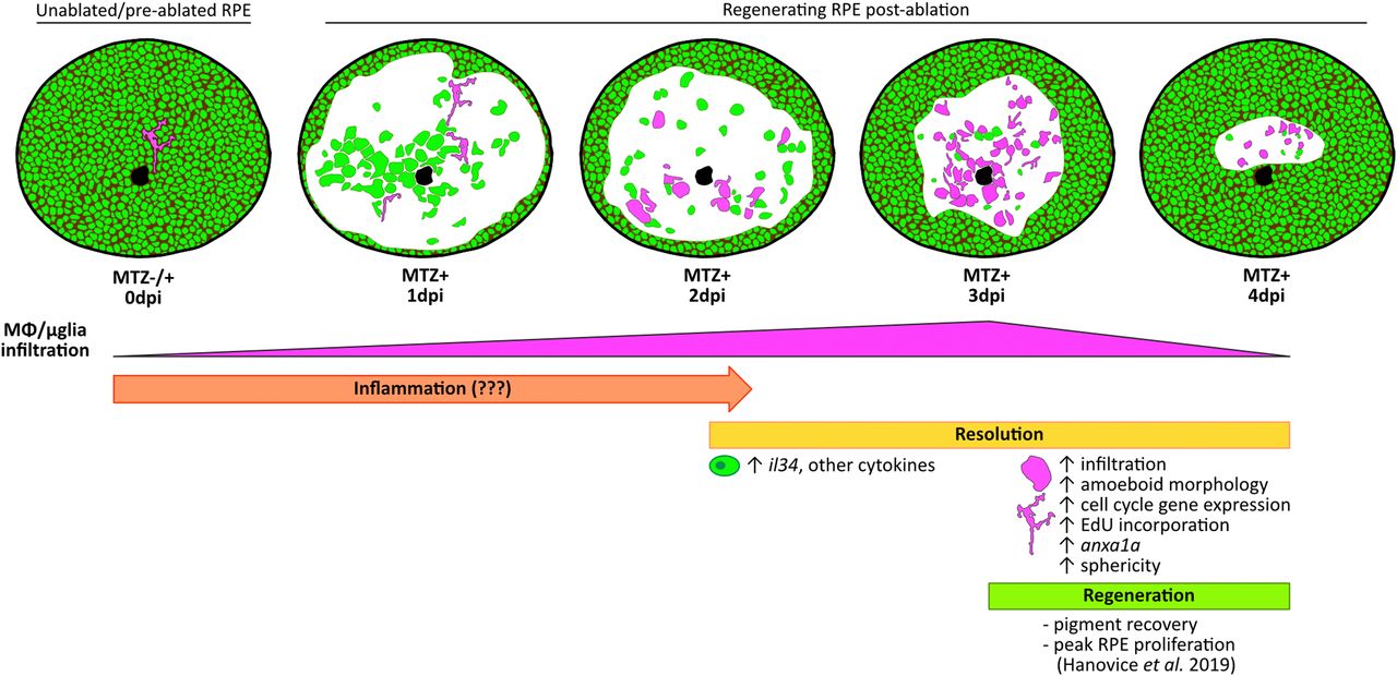
Fig. 8 Phases of immune involvement during RPE regeneration. Schematic showing few ramified MΦs/μglia (magenta) present in the RPE (green) of unablated larvae. Infiltration of MΦs/μglia to the central RPE injury site after ablation begins at 2 dpi, peaks at 3 dpi, and wanes by 4 dpi, representing a time window when inflammation is likely resolved (2 to 4 dpi; yellow). During resolution, MΦs/μglia appear amoeboid in morphology, proliferate and express phagocytosis markers (e.g., anxa1a), and RPE express il34 and other cytokines. Peak RPE layer proliferation and recovery of pigment occurs between 3 to 4 dpi (18). This coupled with the decreased presence of MΦs/μglia in the RPE by 4 dpi may hint to a time window after ablation (3 to 4 dpi; green) when inflammation has been resolved, enabling peak RPE regeneration.
Image
Figure Caption
Acknowledgments
This image is the copyrighted work of the attributed author or publisher, and
ZFIN has permission only to display this image to its users.
Additional permissions should be obtained from the applicable author or publisher of the image.
Full text @ Proc. Natl. Acad. Sci. USA

