
Fig. 5
- ID
- ZDB-FIG-211006-5
- Publication
- Leach et al., 2021 - The immune response is a critical regulator of zebrafish retinal pigment epithelium regeneration
- Other Figures
- All Figure Page
- Back to All Figure Page
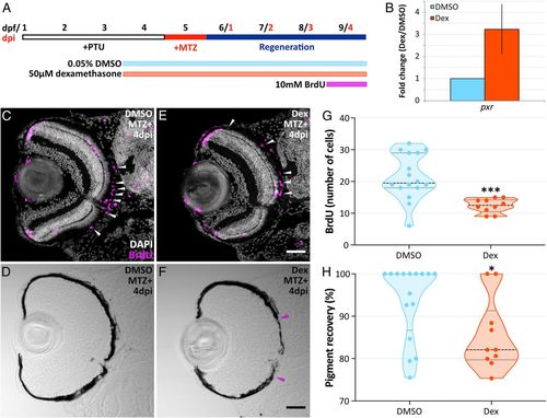
Suppression of inflammation with dexamethasone impairs RPE regeneration. (A) Schematic depicting treatment timeline. (B) Bar graph showing fold change in pxr gene expression from larvae treated with dexamethasone or DMSO for 24 h (4 to 5 dpf). Error bar represents 95% CI. (C–F) Confocal micrographs of transverse sections from 4 dpi MTZ+ DMSO- (C and D) and dexamethasone-treated (E and F) Tg(rpe65a:nfsB-eGFP) eyes. (C and E) White arrowheads highlight BrdU+ cells (magenta) in the RPE layer. White (DAPI) labels nuclei. (F) Magenta arrowheads designate edges of the regenerating RPE monolayer. (Scale bars, 40 μm.) (G and H) Violin plots showing a significant decrease in the number of BrdU+ cells in the RPE (G) and the percent recovery of a pigmented monolayer (H) in MTZ+ dexamethasone-treated larvae. Dashed black lines represent the median, and dotted black lines represent quartiles. Statistical information can be found in SI Appendix, Table S12. Dorsal is up; *P value ≤ 0.05; and ***P value ≤ 0.001. PTU = n-phenylthiourea. |