|
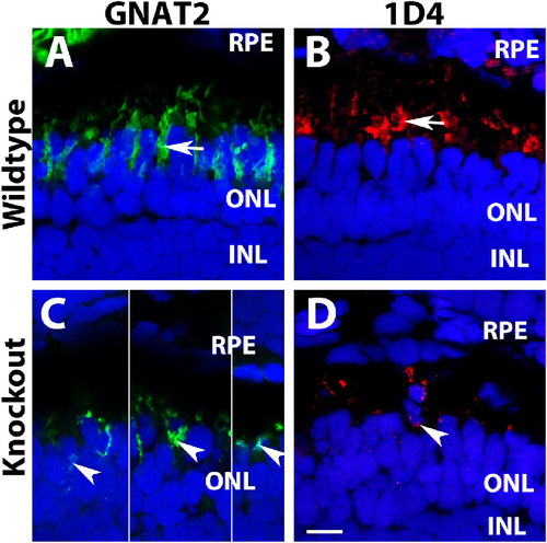
Mislocalization of cone outer segment proteins in tmem216 knockout photoreceptors. Cryosections were immunostained with GNAT2 (green) and 1D4 (red). (A, C) GNAT2 staining for wildtype and tmem216snyR8Δ60 homozygous retina. Similar phenotypes were observed in the tmem216snyΔ175 homozygous retina. GNAT2 was normally found in the cone outer segments (arrow in A) in the wildtype retina. Mislocalization of GNAT2 reactivity to the cone cell body was frequently found in the knockout fish (arrowheads in C). (B, D) 1D4 staining of wildtype and knockout retina. 1D4 reactivity was labeling long double cone outer segments in the wildtype (arrow in B). 1D4 reactivity was frequently found around the cell bodies of the knockout retina (arrowhead, in D). Scale bar in D: 6 µm.
|