|
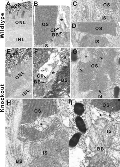
tmem216 knockouts exhibit abnormal outer segment disc morphology. Eyes from 7-dpf wildtype and tmem216snyR8Δ60 zebrafish were processed for transmission EM. (A–D) Wildtype retina. Note that the outer segments with uniform discs. (E–I) tmem216snyR8Δ60 homozygous retina. Although photoreceptors in the tmem216snyR8Δ60 homozygous retina elaborated cilia (asterisk in F), the outer segments of these photoreceptors exhibit multiple defects. Abnormalities manifested as large vacuoles within the outer segment (asterisks in G and H), large vacuoles at the base of the outer segment (asterisks in I), shortened discs (arrows in G), abnormal disc morphology (arrowhead in G). Scale bar in I: 4 µm for A and E; 400 nm for B, D, and F; 200 nm for C, G, H, and I. BB, basal body; CP, ciliary pocket; IS, inner segment; OS, outer segment.
|