
Figure 4.
- ID
- ZDB-IMAGE-200829-68
- Antibodies
- Publication
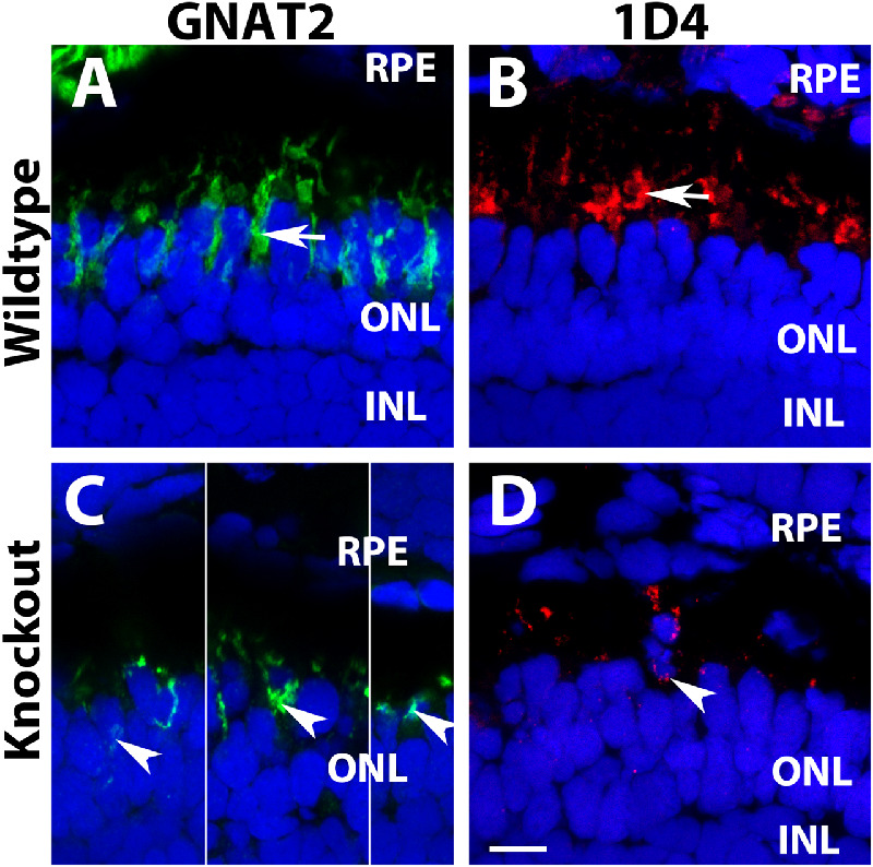
- Liu et al., 2020 - TMEM216 Deletion Causes Mislocalization of Cone Opsin and Rhodopsin and Photoreceptor Degeneration in Zebrafish
- All Figures
- Figures for Liu et al., 2020
|
Figure 4.
Mislocalization of cone outer segment proteins in