|
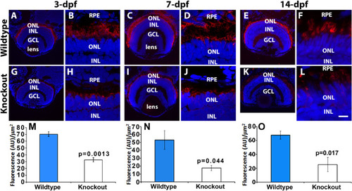
Rod outer segments were reduced in tmem216 knockout zebrafish. Retinal sections were stained with rhodopsin marker, 4D2 (red), and counterstained with DAPI to label nuclei. Knockout images shown represent the tmem216snyΔ175 retina, similar phenotypes were observed in the tmem216snyR8Δ60 homozygous retina. (A–F) Wildtype at 3-, 7-, and 14-dpf, respectively. (G–L) tmem216snyΔ175 homozygous at 3-, 7-, and 14-dpf, respectively. (M–O) 4D2 immunofluorescence intensity was significantly reduced at 3-, 7-, and 14-dpf in tmem216snyΔ175 homozygous retina (n = 3, Student's t-test). Scale bar in L: 55 µm for A and G; 110 µm for C, I, E, and K; 6 µm for B, H, D, J, F, and L.
|