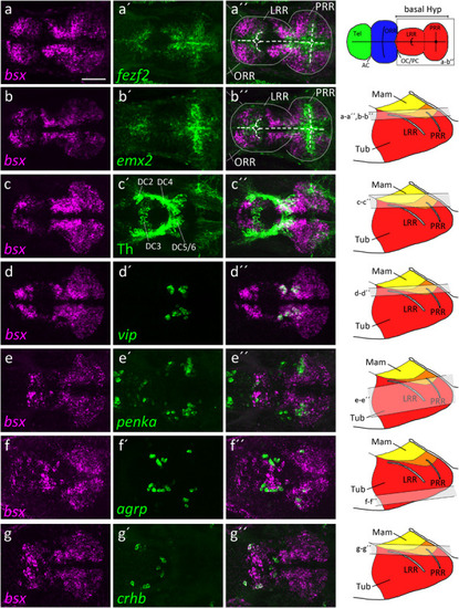
bsx expression within the bHyp in relation to expression of neural progenitor markers and neuropeptidergic genes. Dorsal view of confocal sections of zebrafish embryos at 2 dpf (a–b′′), 3 dpf (c–e′′) or 4 dpf (f–g′′) after double-fluorescent whole-mount in situ hybridization using probes as indicated. Maximum intensity Z-projections of 30 (a–b′′,d–d′′), 40 (f–g′′), 50 (c–c′′), or 70 (e–e′′) single confocal planes (1 μm steps) are shown. Schematics on the right show lateral view of the bHyp for 2–4 dpf embryos indicating which planes were selected for Z-projections in the panels indicated. Expression of bsx is detected further away from the ventricle [white dashed line in (a–b′′)] than expression of neuronal progenitor markers (a–b′′). bsx expression colocalizes in neurons expressing Th [anti-Th immunostain; (c–c′′)] or neuropeptidergic transcripts (d–g′′). Scale bar in (a) for all images: 50 μm. Schematics in the top right represents a model of the zebrafish forebrain highlighting the ventricular recesses. Anterior at left. For abbreviations see list.
|
