|
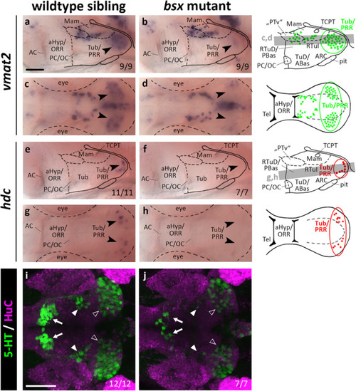
vmat2 expression, hdc expression and 5-HT immunoreactivity in the posterior recess region are reduced or absent in bsx mutant embryos. Lateral (a,b,e,f) and dorsal views (c,d,g,h) of the ventral forebrain in 3 dpf embryos stained by in situ hybridization using probes as indicated. (i,j) Dorsal view of the ventral forebrain in 3 dpf embryos immunostained using 5-HT (green) and HuC antibody (magenta). Images show minimum intensity projections of 40 brightfield focal planes (a–h; 1 μm steps) or maximum intensity projections of 40 confocal planes (i,j; 1 μm steps). Scale bars in (a) for (a–h) and in (i) for (i) and (j): 50 μm. Anterior at left. Schematics to the right show location of vmat2 (green) and hdc (red) expressing cells as observed in stainings and indicate the focal planes which are shown in dorsal view pictures and schematics. Cells with Bsx-dependent expression are circled. Numbers n (phenotype shown)/n (total analyzed) as indicated.
|