FIGURE

Figure 6—figure supplement 1.

ID
ZDB-FIG-200612-23
Publication
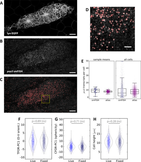
Hartmann et al., 2020 - An image-based data-driven analysis of cellular architecture in a developing tissue
Other Figures
All Figure Page
Back to All Figure Page
Figure 6—figure supplement 1.

Spot Detection and Cell Shape Embedding for <italic>pea3</italic> smFISH Data.

(A–C) Maximum z-projections of a two-color sample showing the lyn-EGFP membrane marker (A), the pea3 smFISH probe (B), and the results of automated spot detection with red rings denoting detection events (C). Scale bars: 10 µm. (D) Zoomed view of the region in the yellow box in (C). Scale bar: 2 µm. (E) pea3 smFISH spot counts for each cell, both from measured data (blue) and from predictions across the entire atlas dataset (purple). The left shows averages across primordia, which closely match those reported previously based on a different spot counting method (Durdu et al., 2014). The individual cell counts on the right show that there is a long tail of cells with extremely high counts, which as one would expect is not captured in the SVR predictions. (F–H) Comparisons of three important cell shape variables between fixed smFISH samples and live samples (here the set of live samples containing only the membrane label; N = 24, n = 2310), showing no significant difference.

Expression Data

Expression Detail
Antibody Labeling
Phenotype Data

Phenotype Detail
Acknowledgments
This image is the copyrighted work of the attributed author or publisher, and ZFIN has permission only to display this image to its users. Additional permissions should be obtained from the applicable author or publisher of the image. Full text @ Elife