
Fig. 2
- ID
- ZDB-FIG-200416-5
- Publication
- Holowiecki et al., 2020 - Pbx4 limits heart size and fosters arch artery formation through partitioning second heart field progenitors and restricting proliferation
- Other Figures
- All Figure Page
- Back to All Figure Page
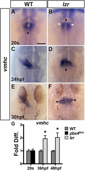
Cardiac fusion and elongation are abnormal in lzr mutants. (A-F) ISH for vmhc in WT and lzr mutant hearts at the 20 s stage, 24 hpf and 30 hpf. Views are dorsal with anterior up. Black arrows in A and B indicate the location of cardiac fusion when forming the cone. White arrows in B indicate anterior aggregates of CMs. Arrow in D indicates the larger ventricular pole. Arrows in F indicate already visible ventricular protrusions. At least 48 embryos per developmental stage were examined and genotyped. Scale bar: 100 µm. (G) RT-qPCR for vmhc expression in WT and Pbx4-depleted embryos at 20 s and in WT and lzr mutants at 36 hpf and 48 hpf. Error bars indicate s.e.m. *P<0.05. |
| Gene: | |
|---|---|
| Fish: | |
| Knockdown Reagents: | |
| Anatomical Terms: | |
| Stage Range: | 20-25 somites to Long-pec |
| Fish: | |
|---|---|
| Observed In: | |
| Stage Range: | Prim-15 to Long-pec |