
FIGURE
Fig. 3
Fig. 3
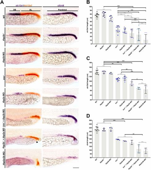
tfap2a and tfap2b function redundantly to activate distal nephron solute transporter signature. (A) Whole-mount in situ hybridization for slc12a1 (DE, purple), slc12a3 (DL, red) and clcnk (pan-distal, purple) at 24 hpf. Black bars indicate wild-type marker domains. Black dots encircle cysts in the duct; black arrowhead indicates incomplete fusion of cloaca. Scale bar: 35 µm. (B-D) Absolute length quantifications of (B) slc12a1, (C) slc12a3 and (D) clcnk. n≥3. Measurements compared by ANOVA. Data are mean±s.d. *P<0.05; **P<0.01; ***P<0.001; N.S., not significant. |
Expression Data
| Genes: | |
|---|---|
| Fish: | |
| Knockdown Reagents: | |
| Anatomical Terms: | |
| Stage: | Prim-5 |
Expression Detail
Antibody Labeling
Phenotype Data
Phenotype Detail
Acknowledgments
This image is the copyrighted work of the attributed author or publisher, and
ZFIN has permission only to display this image to its users.
Additional permissions should be obtained from the applicable author or publisher of the image.
Full text @ Development