
Fig. 6
- ID
- ZDB-FIG-180821-51
- Publication
- Fukui et al., 2018 - Hippo signaling determines the number of venous pole cells that originate from the anterior lateral plate mesoderm in zebrafish
- Other Figures
- All Figure Page
- Back to All Figure Page
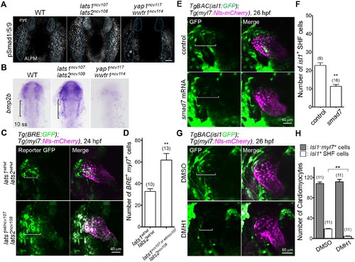
The Hippo signaling pathway functions upstream of the Bmp-dependent signal that is necessary for Isl1-positive SHF formation. (A) Confocal 3D-stack images (at 14 hpf) of embryos immunostained with the anti-pSmad1/5/9 Ab. Blue broken lines indicate the phosphorylated Smad1/5/9-positive cells in the ALPM and eyes. Note that the pSmad1/5/9-positive signals in the ALPM and the eyes are increased and decreased in the embryos of lats1/2 DKO and yap1/wwtr1 DKO, respectively (n = 3). Scale bar indicates 60 μm. (B) WISH analyses at the 10 somite stage (ss) of WT (n = 9), lats1/2 DKO (n = 10), and yap1/wwtr1 DKO (n = 8) embryos, using antisense probe for bmp2b. Square brackets indicate the bmp2b-positive ALPM. (C) Confocal 3D-stack images (at 24 hpf) of the Tg(BRE:GFP);Tg(myl7:Nls-mCherry) embryos carrying lats1wt/wtlats2wt/wt (upper panels) and lats1wt/ncv107lats2ncv108 alleles (bottom panels). Square brackets highlight the GFP-positive myl7-promoter-active cells in the venous pole. Note that the numbers of GFP-positive myl7-promoter-active cells are increased in the venous pole. (D) Quantitative analyses of the numbers of the both BRE-active GFP-positive and myl7-promoter-active mCherry-positive cells in the lats1wt/wtlats2wt/wt embryos and in either the lats1wt/ncv107lats2ncv108 embryos or the lats1/2 DKO embryos. (E) Confocal 3D-stack images (at 26 hpf) of the TgBAC(isl1:GFP);Tg(myl7:Nls-mCherry) control embryos (uninjected, upper panels) and embryos injected with 100 pg of smad7 mRNA (bottom panels). Square brackets indicate the region of isl1-promoter-active GFP-positive SHF cells in the venous pole. (F) Quantitative analyses of the number of isl1-promoter-active SHF cells in the venous pole. Note that overexpression of smad7 mRNA leads to a decrease in the number of isl1-promoter-active SHF cells in the venous pole. (G) Confocal 3D-stack images (at 26 hpf) of the TgBAC(isl1:GFP);Tg(myl7:Nls-mCherry) embryos treated with DMSO (upper panels) or DMH1 (10 μM, lower panels) constructed between 14 hpf and 26 hpf. Square brackets indicate the isl1-promoter-active SHF cells in the venous pole. (H) Quantitative analyses of the number of isl1-promoter-active SHF cells and of the number of both GFP-negative and myl7-promoter-active mCherry-positive cells. Note that DMH1 treatment decreases the number of isl1-promoter-active SHF cells in the venous pole but does not affect the number of GFP-negative and myl7-promoter-active CMs. All images in this figure are in dorsal view, anterior to the top. The confocal 3D-stack images and ISH images are a set of representative images of at least three independent experiments. **p < 0.01. |
| Genes: | |
|---|---|
| Antibody: | |
| Fish: | |
| Condition: | |
| Anatomical Terms: | |
| Stage Range: | 10-13 somites to Prim-5 |
| Fish: | |
|---|---|
| Condition: | |
| Observed In: | |
| Stage Range: | 10-13 somites to Prim-5 |