
Fig. 1
- ID
- ZDB-FIG-180821-37
- Publication
- Fukui et al., 2018 - Hippo signaling determines the number of venous pole cells that originate from the anterior lateral plate mesoderm in zebrafish
- Other Figures
- All Figure Page
- Back to All Figure Page
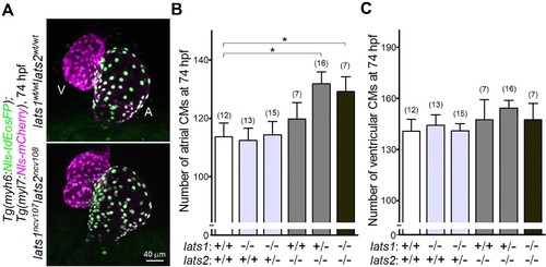
Knockout of lats1/2 genes leads to an increase in the number of atrial, but not ventricular CMs during early development. (A) Confocal 3D-stack images (at 74 hr post fertilization [hpf]) of the Tg(myh6:Nls-tdEosFP);Tg(myl7:Nls-mCherry) embryos with lats1wt/wtlats2wt/wt (top) and lats1ncv107lats2ncv108 alleles (bottom). Atrial (A) and ventricular (V) cardiomyocytes (CMs) are EosFP-positive cells and EosFP-negative mCherry-positive cells, respectively. Ventral view, anterior to the top. (B, C) Quantitative analyses of the number of atrial (B) and ventricular (C) CMs of the embryos at 74 hpf with alleles indicated at the bottom. Plus (+) and minus (–) signs indicate the wt allele and the allele of ncv107 or ncv108 in lats1 or lats2 genes, respectively. The confocal 3D-stack images are a set of representative images of eight independent experiments. In the graphs, the total number of larvae examined in the experiment is indicated on the top of columns unless otherwise described. *p < 0.05. |
| Genes: | |
|---|---|
| Fish: | |
| Anatomical Terms: | |
| Stage: | Protruding-mouth |
| Fish: | |
|---|---|
| Observed In: | |
| Stage: | Protruding-mouth |