
Fig. 2
- ID
- ZDB-FIG-180821-40
- Publication
- Fukui et al., 2018 - Hippo signaling determines the number of venous pole cells that originate from the anterior lateral plate mesoderm in zebrafish
- Other Figures
- All Figure Page
- Back to All Figure Page
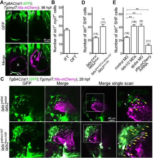
The Hippo signaling pathway is involved in the formation of Isl1-positive SHF cells in the venous pole. (A) Single-scan confocal images (at 96 hpf) of the TgBAC(isl1:GFP);Tg(myl7:Nls-mCherry) embryos. Cells in which both the isl1 and myl7 promoters are active are present in the inflow tract (IFT) cells (arrows) but not in the outflow tract (OFT) cells (square bracket). A, atrium; V, ventricle. Ventral view, anterior to the top. (B) Quantitative analyses of the number of cells with both isl1- and myl7-promoter activities in the IFT and the OFT at 96 hpf (n = 10). (C) Confocal images (at 26 hpf) of the TgBAC(isl1:GFP);Tg(myl7:Nls-mCherry) embryos with the lats1wt/wtlats2wt/wt (upper panels) or lats1wt/ncv107lats2ncv108 alleles (bottom panels). The boxed regions are enlarged in the panels in the fourth (right) column. Yellow arrows indicate both isl1- and myl7-promoter activities in cells in the venous pole. White arrowheads indicate cells with isl1-promoter activity that are in contact with cells in which there is myl7-promoter activity. Confocal 3D-stack images (left two panels) and single-scan images (right two panels). Dorsal view, anterior to the top. (D, E) Quantitative analyses of the number of the isl1-promoter-active SHF cells in the venous pole of the lats1wt/wtlats2wt/wt embryos, the lats1wt/ncv107lats2ncv108 embryos, and the lats1/2 DKO embryos (D), and in embryos shown in Figure 2—figure supplement 1D (E). Both cells with isl1- and myl7-promoter activity and with isl1-promoter activity that were in contact cells with myl7-promoter activity were counted as SHF cells. The confocal 3D-stack images and single-scan (2 μm) images are a set of representative images of at least four independent experiments. **p < 0.01. |
| Genes: | |
|---|---|
| Fish: | |
| Knockdown Reagents: | |
| Anatomical Terms: | |
| Stage Range: | Prim-5 to Day 4 |
| Fish: | |
|---|---|
| Knockdown Reagents: | |
| Observed In: | |
| Stage: | Prim-5 |